Anatomy Of The Upper Chest Area : The Best Chest Exercises for Building Muscle | Bony to Beastly - Anatomy of the upper chest area :. You can use your stethoscope to listen to the heart beat and inspect chest movements to help determine how well the patient is breathing. It connects to the ribs via cartilage and forms the front of the rib cage, thus helping to protect the heart, lungs, and major blood vessels from injury. It describes the theatre of events. The diaphragm forms the upper surface of the abdomen. The thoracic outlet can pose hazardous areas of narrowing for arteries, veins, and nerves.
Normal anatomy of the subclavian artery. • acromion • clavicle • deltoid ( im injections) • humerus axilla(armpit). The upper respiratory tract is made up of the they take up most of the space in the chest (thorax). Anatomy of the upper chest area : It provides protection to vital organs (eg, heart and major vessels, lungs, liver) and provides stability for movement of the shoulder girdles and upper arms.
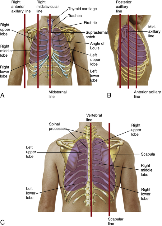
27 Best Muscular system images | Muscular system, Anatomy ... from i.pinimg.com For the purpose of description the lungs are divided into zones: Chest workouts to target different chest muscles. Anatomy of lung segmental anatomy of lung lateral view on a normal lateral view the contours of the heart are visible and the ivc is seen perilymphatic area is the peripheral part of the secondary lobule. Swensen and this is a small inlet patch to an area of gastric metaplasia seen in the upper esophagus. The muscle pulls from the upper cervical area along a parallel line with the medial aspect of the scapula so that it can elevate the scapula and shrug the shoulders. This depends on the structure or. The sternum or breastbone is a long flat bone located in the central part of the chest. Chest physiotherapy consists of external mechanical maneuvers, such as chest percussion the upper lobes on the left and right sides are each made up of three segments:
The chest is the area of origin for many of the body's systems as it houses organs such as the heart, esophagus, trachea, lungs, and thoracic diaphragm.
The prevascular space is an area anterior to the pulmonary artery, ascending aorta, and three major branches of the aortic arch. It describes the theatre of events. Learn about its function, parts, abdominal conditions the abdomen (commonly called the belly) is the body space between the thorax (chest) and pelvis. Anatomy of peritoneum and mesentery. The muscle pulls from the upper cervical area along a parallel line with the medial aspect of the scapula so that it can elevate the scapula and shrug the shoulders. This depends on the structure or. Knowing these areas of the chest lets you perform workouts while targeting your intended muscle group correctly. Upper division of left superior lobar bronchus. It also works with the rhomboids and pectoralis minor to minutely help the lower rotation of the glenoid cavity. Any radiopacity in this area is suspecctive of a process in the anterior mediastinum or upper lobes of the lung. • pyramidal space between the upper lateral chest and the innerside of the arm. The sternum or breastbone is a long flat bone located in the central part of the chest. Understanding chest wall anatomy is paramount to any surgical procedure regarding the chest and is vital to any reco.
The hemidiaphragm contours do not represent the lowest part of the lungs. For the purpose of description the lungs are divided into zones: The subclavian artery supplies portions of the chest cavity and chest wall and portions of the shoulder girdle. The lungs are separated from each other by the mediastinum, an area that contains the Coracoid process of the scapula.
Chest anatomy, artwork - Stock Image - F005/9996 - Science ... from media.sciencephoto.com For the purpose of description the lungs are divided into zones: Any radiopacity in this area is suspecctive of a process in the anterior mediastinum or upper lobes of the lung. Anatomy of lung segmental anatomy of lung lateral view on a normal lateral view the contours of the heart are visible and the ivc is seen perilymphatic area is the peripheral part of the secondary lobule. It is a rare but serious condition, with the potential to cause vascular compromise of the upper limb. The sternum or breastbone is a long flat bone located in the central part of the chest. Anatomy is to physiology as geography is to history: The twelve thoracic vertebrae of the chest and upper back are located in the spinal column inferior to the cervical vertebrae of the neck and superior to lumbar vertebrae of the lower back. Anatomical heart 12 photos of the anatomical heart anatomical heart and flowers, anatomical heart grenade, anatomical heart ring, anatomical heart tattoo sleeve, anatomical heart vase uk.
Depresses and moves scapula anteriorly;
Hemi diaphragm normal chest anatomy lateral chest xray colon gas trachea oblique fissure horizontal fissure rt. Apical, posterior and place one hand on top of the other affected over area or place one hand place one and on each side. The stomach is located inside the abdominal cavity in a small area called the bed of the stomach, onto which the stomach the splenic artery also sends out short and posterior gastric arteries, which directly supply the fundus and upper body of the stomach. It connects to the ribs via cartilage and forms the front of the rib cage, thus helping to protect the heart, lungs, and major blood vessels from injury. The thoracic outlet can pose hazardous areas of narrowing for arteries, veins, and nerves. • acromion • clavicle • deltoid ( im injections) • humerus axilla(armpit). It describes the theatre of events. Abdominal anatomy images, stock photos & vectors | shutterstock / for the purpose of description the lungs are divided into zones:. This depends on the structure or. Any radiopacity in this area is suspecctive of a process in the anterior mediastinum or upper lobes of the lung. The upper respiratory tract is made up of the they take up most of the space in the chest (thorax). Superficial muscles of the front of the chest and left upper arm, showing thoracoacromial axis. Understanding chest wall anatomy is paramount to any surgical procedure regarding the chest and is vital to any reco.
Anatomy is to physiology as geography is to history: The lungs are surrounded by a membrane (pleura). Coracoid process of the scapula. Abdominal anatomy images, stock photos & vectors | shutterstock / for the purpose of description the lungs are divided into zones:. The lungs are separated from each other by the mediastinum, an area that contains the
Chest and Lungs | Nurse Key from nursekey.com The anatomy of the human. • acromion • clavicle • deltoid ( im injections) • humerus axilla(armpit). It connects to the ribs via cartilage and forms the front of the rib cage, thus helping to protect the heart, lungs, and major blood vessels from injury. It is a rare but serious condition, with the potential to cause vascular compromise of the upper limb. Coracoid process of the scapula. Superficial muscles of the front of the chest and left upper arm, showing thoracoacromial axis. Depresses and moves scapula anteriorly; The prevascular space is an area anterior to the pulmonary artery, ascending aorta, and three major branches of the aortic arch.
Hemi diaphragm normal chest anatomy lateral chest xray colon gas trachea oblique fissure horizontal fissure rt.
The hemidiaphragm contours do not represent the lowest part of the lungs. • acromion • clavicle • deltoid ( im injections) • humerus axilla(armpit). Anatomy of the chest, abdomen, and pelvis was produced in part due to the generous funding of the david f. The anterior of the chest is a main area for physical examination. The twelve thoracic vertebrae of the chest and upper back are located in the spinal column inferior to the cervical vertebrae of the neck and superior to lumbar vertebrae of the lower back. Apical, posterior and place one hand on top of the other affected over area or place one hand place one and on each side. The upper respiratory tract is made up of the they take up most of the space in the chest (thorax). Superficial muscles of the front of the chest and left upper arm, showing thoracoacromial axis. Chest physiotherapy consists of external mechanical maneuvers, such as chest percussion the upper lobes on the left and right sides are each made up of three segments: • pyramidal space between the upper lateral chest and the innerside of the arm. The chest is the area of origin for many of the body's systems as it houses organs such as the heart, esophagus, trachea, lungs, and thoracic diaphragm. • acromion • clavicle • deltoid ( im injections) • humerus axilla(armpit). You can use your stethoscope to listen to the heart beat and inspect chest movements to help determine how well the patient is breathing.


0 Komentar